You’re in your 30s or 40s feeling fine, but your doctor mentions osteoporosis risk and suddenly you’re wondering if your bones are silently weakening, which is exactly why understanding how a bone density scan DXA explained can transform your approach to long-term health.
Understanding bone density scan
A bone density scan, formally known as dual-energy X-ray absorptiometry or DXA, represents one of the most straightforward yet powerful tools in modern medicine for evaluating skeletal health. Unlike traditional X-rays that simply show bone structure, DXA measures the actual mineral content within your bones, giving clinicians a numerical value that indicates how dense and strong your skeleton really is. Think of it like checking the structural integrity of a building: you’re not just looking at the walls, you’re measuring how solid they are. This measurement becomes particularly relevant when assessing conditions like osteoporosis, where bones gradually lose density and become more fragile. Young adults might not think about bone density yet, but the foundation you build now directly impacts your skeletal health decades from now. DXA scans help identify early warning signs before fractures occur, making them invaluable for preventive health strategies.
The science of DXA technology
DXA technology operates on a clever principle: it fires two distinct X-ray beams at different energy levels through your body, and by measuring how much radiation each beam absorbs, the machine calculates bone mineral density with remarkable precision. Here’s where it gets interesting: different tissues absorb X-rays differently. Bone absorbs more radiation than soft tissue, and the denser the bone, the more absorption occurs. The computer compares the absorption patterns between the two energy levels to eliminate interference from surrounding soft tissue like muscle and fat, giving you a pure measurement of bone density alone. This dual-beam approach is what makes DXA superior to older single-beam technology. Researchers have refined this method over decades, and modern DXA machines can detect density changes as small as 1 to 2 percent, making them sensitive enough to track bone health changes over time. For young adults, understanding this technology demystifies what happens during the scan and explains why the results are so reliable.
How DXA scans are performed
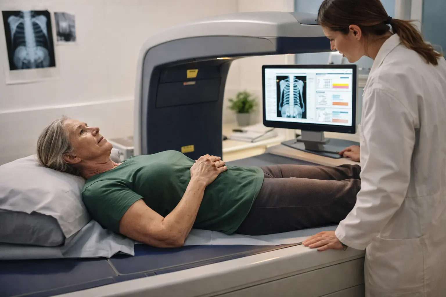
The actual DXA scan procedure is refreshingly simple and non-invasive, typically taking between 10 and 30 minutes depending on which bones are being measured. You’ll lie flat on a padded scanning table in comfortable clothing, and the technician will position you so the scanner arm can pass over the areas being assessed, usually the hip, spine, and sometimes the forearm. The scanner arm moves slowly across your body, emitting low-dose X-rays while a detector on the opposite side captures the radiation that passes through. You won’t feel anything during this process, and there’s no pain or discomfort involved. The key is remaining still so the images stay sharp and accurate. Once the scan completes, a computer analyzes the captured images, calculating bone mineral density at multiple points and comparing your results to reference databases of healthy young adults and people your age and gender. This comparison generates your T-score and Z-score, which your healthcare provider will interpret to assess your bone health status and fracture risk.
- Lie on the scanning table in comfortable clothing while the technician positions you for optimal imaging.
- Remain still as the scanner arm passes over your body, emitting low-dose X-rays to capture bone density data.
- Allow the computer to analyze your images and generate your bone mineral density scores and comparison results.
- Consult with your healthcare provider to understand your T-score, Z-score, and what they mean for your individual bone health.
🔬 Science-backed benefits in 2 minuteschoose where to begin:
Interpreting DXA results
Your DXA results arrive as a T-score, which compares your bone mineral density to that of a healthy 30-year-old adult of your same sex, essentially showing how your bones stack up against peak bone mass. A T-score of -1 or higher is considered normal, meaning your bones are as dense as or denser than the reference standard. Between -1 and -2.5 indicates osteopenia, a condition where bone density is lower than normal but not yet at the osteoporosis threshold, think of it as a yellow light rather than a red one. A T-score below -2.5 indicates osteoporosis, where bone density has declined significantly and fracture risk increases substantially. You’ll also receive a Z-score, which compares your results to people your exact age and sex, providing context about whether your bone density is typical for your demographic. For young adults, even if your T-score is normal now, understanding these numbers helps you make informed decisions about lifestyle factors like exercise, nutrition, and calcium intake that influence bone health trajectories.
Benefits and limitations of DXA
DXA scans offer tremendous value as a non-invasive, low-radiation screening tool that provides objective, quantifiable data about bone health without requiring any injections or contrast dyes. The procedure is quick, painless, and widely available at most hospitals and imaging centers, making it accessible for routine screening. Results are highly reproducible, meaning if you get scanned twice, the measurements will be nearly identical, allowing clinicians to track changes over months or years with confidence. However, DXA does have limitations worth understanding. It measures bone density but not bone quality or microarchitecture, so two people with identical T-scores might have different fracture risks based on bone structure. The scan can be less accurate in people with spinal deformities, severe arthritis, or metal implants like hip replacements, since these interfere with X-ray absorption. Additionally, DXA doesn’t account for falls, balance problems, or muscle strength, all of which significantly influence actual fracture risk. For young adults, recognizing these limitations means DXA results should inform but not solely determine your health decisions.
Future applications of DXA
The future of bone density assessment is moving beyond simple mineral measurement toward more sophisticated analysis of bone quality and structure. Researchers are developing three-dimensional imaging capabilities that would allow DXA machines to evaluate bone microarchitecture, the intricate lattice-like structure within bones that determines strength independent of density alone. Some institutions are exploring how DXA data combined with artificial intelligence algorithms could predict fracture risk more accurately than traditional scoring systems. Advanced software is being refined to assess trabecular bone score, a measure of bone texture that captures information about the internal structure of bones, providing additional insight into fracture risk. There’s also growing interest in using DXA technology to evaluate bone health in younger populations, potentially identifying individuals at risk for accelerated bone loss due to medications, hormonal factors, or lifestyle choices. For young adults, these advancements suggest that future bone health screening will become even more personalized and predictive, potentially allowing for earlier intervention before significant bone loss occurs.
DXA, or dual-energy X-ray absorptiometry, serves as a cornerstone tool for assessing bone health by precisely measuring bone mineral density through two distinct X-ray beams. The procedure involves lying still while a scanner captures images that a computer analyzes to generate T-scores and Z-scores, which are then compared to reference databases to determine osteoporosis risk and overall skeletal health status. While DXA offers significant benefits as a non-invasive, low-radiation screening method, it has limitations in certain populations and measures density rather than bone quality. Emerging technologies promise more sophisticated assessments of bone microarchitecture and structure, potentially revolutionizing how we predict and prevent fractures in the future.
How often should I get a DXA scan?
Screening frequency depends on individual risk factors including age, sex, family history, medications, and lifestyle factors. Young adults without significant risk factors typically don’t need routine screening, but those with risk factors like prolonged corticosteroid use, hormonal imbalances, or family history of osteoporosis should consult their healthcare provider about appropriate screening intervals. Generally, if you have normal bone density, rescanning every two to three years is common, though some providers recommend longer intervals.
Are DXA scans safe?
DXA scans are considered very safe, emitting radiation doses significantly lower than standard chest X-rays, roughly equivalent to a few days of natural background radiation exposure. The benefits of early bone health detection and fracture risk assessment substantially outweigh the minimal radiation risk, particularly for individuals with risk factors for osteoporosis. Pregnant individuals should inform their technician, though DXA is not typically performed during pregnancy as a precaution.
Others also read:
Disclaimer: This article is for informational purposes only and is not a substitute for professional medical advice. Always consult a healthcare professional for personal guidance.
This article has been prepared and reviewed by the GlobalHealthBeacon editorial team and is based on current medical research and published scientific literature available in 2026. It provides structured, evidence-based information to support informed health decisions.
