You’re noticing your posture isn’t quite what it used to be, or maybe you’ve heard one too many stories about friends breaking bones from minor falls, and now you’re wondering if your skeleton is silently weakening without you knowing it, which is exactly why understanding bone density scan dxa explained has become essential for taking real control of your bone health.
Understanding bone density scan dxa
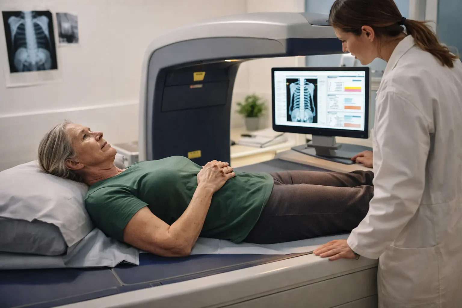
A bone density scan DXA, or dual-energy X-ray absorptiometry, is a straightforward medical test that measures how much mineral is packed into your bones. Think of it like a report card for your skeletal system. The scan uses two different energy levels of X-rays to calculate the exact amount of bone mineral in specific areas, usually your hip, spine, and sometimes your forearm. For women, this test becomes increasingly important as you age because hormonal changes, especially after menopause, can cause bones to lose density rapidly. The procedure itself is completely painless and non-invasive. You simply lie on a padded table while a scanner arm passes over your body, taking detailed measurements. Many women find it less intimidating than they expected. The real value here is that DXA gives your doctor concrete numbers about your fracture risk, which means you can move from guessing about your bone health to having actual data that guides real decisions.
- DXA scan evaluates the strength and thickness of your bones, helping doctors determine if you have osteoporosis or low bone density.
- The results of the DXA scan are compared to the bone density of a healthy young adult, called T-score, and someone of your own age, called Z-score.
- The test is quick, typically taking around 10-30 minutes, and involves lying down on a table while a scanner passes over your body.
Preparing for a bone density scan dxa
Getting ready for your DXA scan is simpler than you might think, but a few details matter. Start by stopping calcium supplements at least 24 hours before your appointment because they can create false readings on the scan. On the day of your test, wear comfortable, loose-fitting clothing without metal components like zippers, buttons, underwire bras, or metal jewelry. These items can interfere with the imaging and may require you to change into a gown, which wastes time. If you’ve had any recent imaging studies involving contrast dye or barium, let your technician know because these can also affect results. Some facilities ask you to remove your shoes and any metal accessories before entering the scanning room. It’s worth calling ahead to ask about their specific requirements. Arrive a few minutes early to complete any paperwork so you’re not rushed. Many women appreciate knowing that the technician will be right there with you the entire time, and you can ask questions anytime during the process.
📘 Fix your day in under 2 minuteschoose where to begin:
During the dxa scan
When you arrive for your scan, the technician will position you on a padded table and explain exactly what will happen. You’ll need to lie very still while the scanning arm moves slowly over your body, usually starting at your feet and working upward. The arm doesn’t touch you, and you won’t feel anything during the scan itself. The technician may ask you to hold your breath briefly during certain measurements to ensure clarity. The entire process typically takes 10 to 30 minutes depending on which areas are being scanned. If you’re having just a hip and spine scan, it’s usually on the shorter end. The radiation exposure is remarkably low, roughly equivalent to the natural radiation you’d receive on a cross-country airplane flight. After the scan is complete, you can immediately return to your normal activities with no recovery time needed. Your doctor will review the images and measurements, then schedule a follow-up appointment to discuss what the results mean for your specific situation and any next steps you should consider.
Interpreting dxa results
Your DXA results will come back with two key numbers: your T-score and Z-score. The T-score compares your bone density to that of a healthy 30-year-old woman at peak bone mass, while the Z-score compares you to women your own age and body size. A T-score of minus 1.0 or higher is considered normal. Between minus 1.0 and minus 2.5 is classified as low bone mass or osteopenia, which means your bones are weaker than ideal but not yet at the osteoporosis stage. A T-score below minus 2.5 indicates osteoporosis. Understanding these numbers helps you and your doctor make informed decisions about treatment and lifestyle changes. For example, if your results show osteopenia, your doctor might recommend increased calcium and vitamin D intake, weight-bearing exercise, and possibly a follow-up scan in one or two years rather than immediate medication. If you have osteoporosis, medication options exist that can slow or stop bone loss. Your doctor will explain your specific results in context of your age, family history, and other risk factors, creating a personalized plan rather than a one-size-fits-all approach.
Maintaining bone health
Strong bones don’t happen by accident, especially for women navigating hormonal changes throughout life. Start with nutrition: aim for 1,000 to 1,200 milligrams of calcium daily from sources like dairy, leafy greens, fortified plant milks, or supplements if needed. Vitamin D is equally critical because your body can’t absorb calcium without it. Get 600 to 800 IU daily from fatty fish, egg yolks, or supplements, and spend time in sunlight when possible. Weight-bearing exercises like walking, jogging, dancing, or strength training are powerful bone builders because they force your muscles to pull on your bones, stimulating them to stay strong. Aim for at least 150 minutes of moderate activity weekly. Avoid smoking completely because it accelerates bone loss significantly. Limit alcohol to one drink daily or less. If you’re approaching or in menopause, discuss hormone therapy options with your doctor, as estrogen plays a major role in bone density. Regular DXA rescans, typically every one to two years depending on your results, help you track progress and adjust your approach as needed.
A bone density scan DXA provides crucial information about your bone health, helping to detect osteoporosis and guide preventive measures. Understanding the process, preparation, and interpretation of DXA results is key to maintaining strong and healthy bones.
Is a DXA scan safe?
Yes, a DXA scan is safe and non-invasive, exposing you to low levels of radiation equivalent to a cross-country flight. It is an essential tool for assessing bone health and preventing osteoporosis.
How often should I have a DXA scan?
The frequency of DXA scans depends on your risk factors for osteoporosis. Generally, women over 65 or men over 70, and those with significant risk factors should have a DXA scan every two years.
Others also read:
Disclaimer: This article is for informational purposes only and is not a substitute for professional medical advice. Always consult a healthcare professional for personal guidance.
This guide has been prepared and reviewed by the GlobalHealthBeacon editorial team and reflects current medical research as of 2026. It provides structured, evidence-based information to support informed health decisions.
