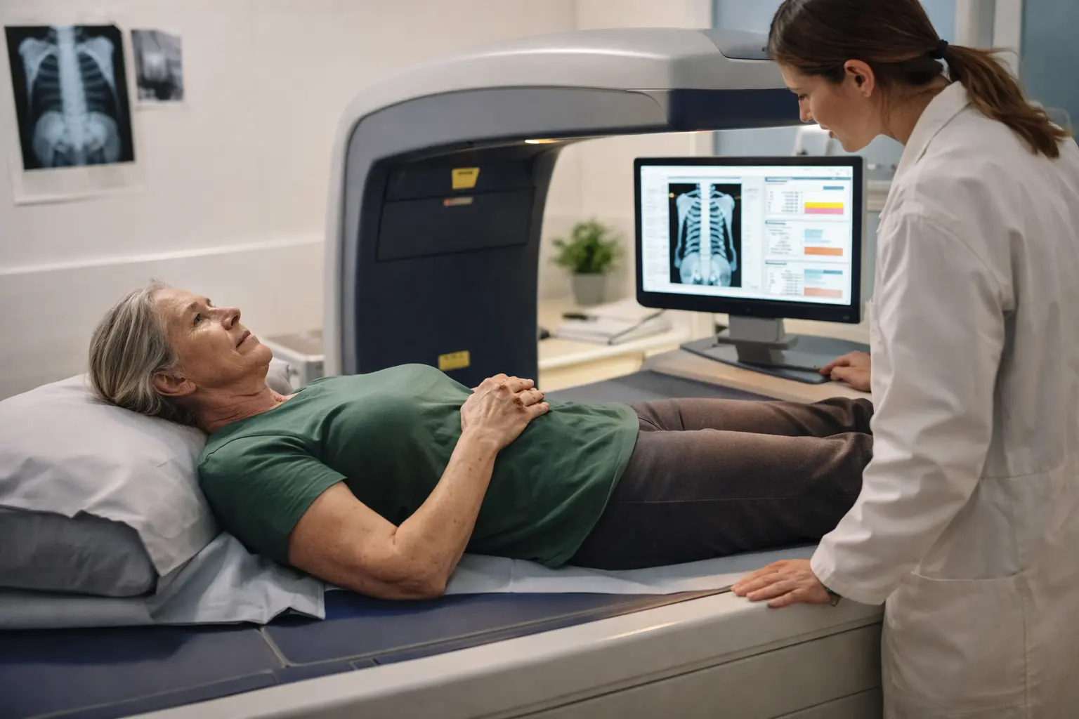
You’re staring at those DXA results and feeling completely lost, wondering if those numbers mean your bones are actually failing you, but here’s what you need to know about bone density scan DXA explained so you can finally stop worrying and start understanding what’s really happening inside your skeleton.
Understanding bone density DXA scan
A bone density scan, formally known as Dual-Energy X-ray Absorptiometry or DXA, works like a specialized camera that photographs the mineral content living inside your bones. Think of it this way: your bones aren’t solid blocks but rather a complex lattice structure, similar to a honeycomb, filled with minerals like calcium and phosphorus. Over time, especially for women navigating menopause or aging, this honeycomb can develop larger spaces, making bones more fragile. The DXA scan measures exactly how tightly packed these minerals are in your skeletal system. This non-invasive test has become the gold standard for assessing fracture risk and diagnosing conditions like osteoporosis, which affects millions of women worldwide. By understanding your bone density now, you’re essentially getting a preview of your skeletal future, allowing you to make informed decisions about your health before problems develop.
How DXA scan works
Imagine lying on a padded table while two different energy X-ray beams pass through your body, each one penetrating bone differently based on mineral density. The scanner measures how much radiation each beam absorbs, and denser bones absorb more radiation. Your results come back as T-scores and Z-scores, two different comparison tools that tell different stories. A T-score compares your bone density to a healthy 30-year-old woman’s peak bone mass, essentially asking how you measure up to optimal bone strength. A Z-score, meanwhile, compares you to women your exact age and ethnicity, answering the question of whether you’re typical for your demographic. Lower scores in either category indicate lower bone density. The scan typically focuses on your hip, spine, or forearm because these areas are most vulnerable to fractures. The entire process takes about 10 to 30 minutes, involves minimal radiation exposure, and provides remarkably detailed information about your skeletal health.
Interpreting DXA results
Your DXA results fall into distinct categories that paint a clear picture of your bone health status. A normal T-score sits above negative 1, meaning your bones are healthy and strong with adequate mineral density. Osteopenia, the middle ground, shows a T-score between negative 1 and negative 2.5, indicating mild bone weakening that warrants attention but doesn’t necessarily mean fractures are imminent. Osteoporosis, the most serious category, presents a T-score below negative 2.5, signaling severely low bone density where fracture risk becomes significantly elevated. Z-scores provide age-specific context, helping your doctor understand whether your bone loss is typical for your age group or accelerated. Consider Sarah, a 58-year-old woman who received a T-score of negative 1.8, placing her in the osteopenia range. While concerning, this score gave her time to implement preventive strategies before progression to osteoporosis. Understanding these categories transforms abstract numbers into actionable health information.
- Review your T-score and Z-score results carefully, noting which category your bone density falls into and what that means for your fracture risk profile.
- Schedule a conversation with your healthcare provider to discuss your specific results, your personal risk factors, and what these numbers mean in the context of your overall health history.
- Work with your provider to develop a personalized plan that may include dietary changes, exercise modifications, medication if appropriate, or lifestyle adjustments to stabilize or improve your bone density over time.
🔬 Science-backed benefits in 2 minuteschoose where to begin:
Importance of DXA scan
DXA scans serve as an early warning system for bone loss, catching problems before they become serious fractures that could change your life. A woman who fractures her hip at 70 might lose independence, mobility, and quality of life, but that same woman screened at 55 with a DXA scan could prevent that outcome entirely through early intervention. Regular DXA scans create a timeline of your bone density, showing whether you’re stable, improving, or declining over months and years. This tracking ability is invaluable because bone loss often happens silently without symptoms until a fracture occurs. If you start medication or lifestyle changes to strengthen your bones, a follow-up DXA scan in one to two years reveals whether your interventions are actually working. For women in midlife transitions, post-menopausal years, or with family histories of osteoporosis, periodic DXA screening becomes an essential tool for maintaining independence and preventing the cascade of health complications that can follow a major fracture.
Risk factors and prevention
Your bone density isn’t fixed by genetics alone; it’s influenced by a complex interplay of age, gender, family history, diet, hormones, and lifestyle choices. Women face particular risk because estrogen, which helps maintain bone density, drops dramatically during menopause, accelerating bone loss. A woman might lose up to 20 percent of her bone density in the five to seven years following menopause. Beyond hormones, inadequate calcium and vitamin D intake weakens your skeletal foundation since these minerals are literally the building blocks of bone. Weight-bearing exercise like walking, dancing, or strength training stimulates your bones to maintain density, while sedentary lifestyles allow bone loss to accelerate. Smoking and excessive alcohol consumption actively damage bone-building cells and reduce calcium absorption. Consider Maria, a 52-year-old woman who started strength training three times weekly, increased her calcium intake to 1,200 milligrams daily, and ensured adequate vitamin D levels. Within two years, her follow-up DXA scan showed stabilized bone density instead of the decline her doctor had predicted. Prevention truly works when you understand the mechanisms driving bone loss.
Consulting a specialist
Your primary care doctor can order a DXA scan and interpret basic results, but a bone health specialist like an endocrinologist or rheumatologist brings deeper expertise to complex situations. If your DXA results show significant bone loss, if you’ve experienced unexplained fractures, or if you have multiple risk factors, specialist consultation becomes valuable. These doctors understand nuanced treatment options, can identify secondary causes of bone loss you might not have considered, and can personalize prevention strategies based on your complete health picture. They might recommend specific medications like bisphosphonates, hormone therapy considerations, or specialized exercise programs designed to maximize bone strength. A specialist can also help you navigate the decision-making process around treatment, weighing benefits against potential side effects based on your individual circumstances. Think of them as your bone health advocates who can answer complex questions and guide you through decisions that might feel overwhelming. Whether you choose specialist care or work with your primary doctor, the key is having someone who takes your bone health seriously and helps you create a sustainable plan for maintaining strong bones throughout your life.
DXA scans provide crucial insights into your bone health by measuring bone density and identifying conditions like osteoporosis. Understanding your DXA results can guide preventive strategies to maintain strong and healthy bones.
Are DXA scans safe?
DXA scans are safe, non-invasive, and emit low levels of radiation, equivalent to a few days of natural background radiation exposure.
How often should I get a DXA scan?
The frequency of DXA scans depends on your age, risk factors, and previous results. Generally, postmenopausal women and men over 50 should undergo DXA scans every 1-2 years.
Others also read:
Disclaimer: This article is for informational purposes only and is not a substitute for professional medical advice. Always consult a healthcare professional for personal guidance.
This article has been prepared and reviewed by the GlobalHealthBeacon editorial team and is based on current medical research and published scientific literature available in 2026. It provides structured, evidence-based information to support informed health decisions.
