You’re worried about your bones breaking, you’ve heard the term DXA thrown around by doctors, and honestly, you have no idea what it actually measures or why it matters for your fracture risk, so let’s break down exactly how a bone density scan DXA explained actually works and what your results really mean.
Understanding bone density scan (DXA)
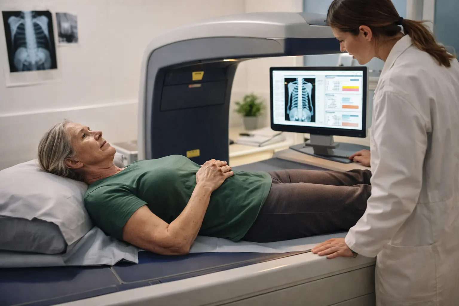
A DXA scan, also called dual-energy X-ray absorptiometry, works by sending two different energy X-ray beams through your bones to measure how much mineral content they contain. Think of it like taking a snapshot of your skeleton’s structural integrity. The machine compares the amount of X-rays that pass through your bones versus soft tissue, giving doctors a precise measurement of bone mineral density. The procedure takes about 10 to 30 minutes depending on which bones are being scanned, typically your hip, spine, or forearm. You lie flat on a padded table while an arm moves slowly over the area being measured. There’s no pain, no injections, and no radiation exposure worth worrying about. The amount of radiation you receive is roughly equivalent to a few hours of natural background radiation from the environment. For young adults, DXA scans are often recommended if you have risk factors like family history of osteoporosis, certain medications that affect bone health, or previous fractures from minor falls.
Importance of bone density in fracture prediction
Your bones are living tissue constantly being broken down and rebuilt, a process called bone remodeling. When bone density decreases, it means there’s less mineral content packed into the same space, making bones more fragile and susceptible to breaking. Imagine a sponge with larger holes versus one with tighter, more densely packed holes. The denser sponge is stronger and less likely to crumble under pressure. Bone density measurements from DXA scans are compared against reference standards to calculate your T-score, which tells you how your bone density compares to a healthy young adult. A T-score of negative 1 or higher is considered normal, between negative 1 and negative 2.5 is classified as osteopenia (low bone mass), and below negative 2.5 indicates osteoporosis. For young adults, maintaining or improving bone density now can significantly reduce fracture risk later in life. Studies show that bone density accounts for roughly 70 percent of bone strength variation, making it a critical predictor of fracture likelihood.
How DXA predicts fractures
DXA predicts fracture risk by establishing a baseline measurement and comparing it against established clinical thresholds developed from large population studies. When your results come back, they’re not just a single number but a comprehensive picture of your skeletal health. The scan measures bone density at multiple sites, and each location tells a different story. Hip measurements are particularly predictive of hip fracture risk, while spine measurements correlate with vertebral fracture risk. Beyond the T-score, doctors also calculate your 10-year fracture probability using tools like FRAX, which incorporates your DXA results along with other risk factors such as age, body weight, smoking status, and alcohol consumption. A young adult with a low T-score might still have a relatively low 10-year fracture probability if other risk factors are minimal. Conversely, someone with normal bone density but multiple other risk factors might warrant preventive intervention. The predictive power of DXA lies in its ability to identify people at higher risk before a fracture occurs, allowing for early intervention through lifestyle modifications, dietary changes, or medication when appropriate.
- Schedule a DXA scan with your healthcare provider if you have risk factors for low bone density, and prepare by wearing comfortable clothing without metal fasteners.
- Review your results with your doctor, understanding your T-score and 10-year fracture probability to grasp your individual risk level.
- Implement recommended interventions such as weight-bearing exercise, adequate calcium and vitamin D intake, and smoking cessation if applicable.
- Follow up with repeat DXA scans as advised by your healthcare provider, typically every 1 to 2 years, to monitor changes in bone density over time.
🔬 Science-backed benefits in 2 minuteschoose where to begin:
Benefits of DXA in fracture prevention
DXA scans serve as an early warning system for bone health problems before they become serious. By identifying low bone density in younger years, you have the opportunity to take action before fractures occur. The scan helps healthcare providers make informed decisions about whether preventive treatment is necessary, potentially avoiding unnecessary medication for people at low risk while ensuring high-risk individuals receive appropriate care. For young adults, DXA results can motivate lifestyle changes like starting a consistent exercise routine or improving nutrition, changes that benefit overall health beyond just bone strength. The scan also provides a baseline for monitoring the effectiveness of interventions over time. If you start taking bone-strengthening medication or increase your exercise routine, follow-up DXA scans can objectively measure whether these changes are working. This feedback loop is powerful because it shows you concrete evidence of improvement, reinforcing healthy behaviors. Additionally, DXA results can inform decisions about other health matters. For instance, certain medications used for other conditions might affect bone density, and knowing your baseline helps doctors choose the safest options for you.
Limitations of DXA in fracture risk assessment
While DXA is valuable, it doesn’t tell the whole story about fracture risk. Bone density accounts for only about 70 percent of bone strength variation, meaning other factors significantly influence whether bones will break. Bone quality, which includes factors like bone microarchitecture and mineralization patterns, isn’t directly measured by DXA but affects fracture risk substantially. Two people with identical T-scores can have very different fracture risks depending on their bone quality. Age is another critical factor that DXA doesn’t fully capture. A 25-year-old and a 75-year-old with the same bone density have vastly different fracture risks because aging affects bone quality and healing capacity. Fall risk is perhaps the most obvious limitation. You could have perfect bone density, but if you fall frequently due to balance problems, vision issues, or medication side effects, your fracture risk remains high. DXA also struggles with certain populations. In younger adults, bone density is often normal even when other risk factors are present, potentially giving false reassurance. Additionally, DXA can’t distinguish between different types of bone loss or predict which specific bones are most vulnerable to fracture.
Research evidence on DXA and fracture prediction
Large-scale studies have consistently shown that DXA measurements correlate with fracture risk, particularly in postmenopausal women and elderly populations where bone loss accelerates. The Fracture Risk Assessment Tool (FRAX) was developed by the World Health Organization based on data from over 60,000 individuals across multiple countries, validating that DXA results combined with clinical risk factors provide meaningful fracture risk prediction. Research demonstrates that for every standard deviation decrease in bone density, fracture risk roughly doubles, though this relationship varies by age and skeletal site. Studies in younger populations show more nuanced results. Young adults with low bone density don’t automatically have high fracture risk if they lack other risk factors, suggesting that age-specific interpretation of DXA results is crucial. Recent research has also highlighted the importance of bone turnover markers and other biomarkers alongside DXA measurements for more complete risk assessment. Meta-analyses confirm that DXA is one of the best non-invasive tools available for fracture risk prediction, but its predictive value improves significantly when combined with clinical judgment and consideration of individual circumstances.
DXA scans provide valuable objective measurements of bone mineral density that help identify individuals at higher risk for fractures, particularly when results are interpreted alongside other clinical factors. Understanding your DXA results empowers you to make informed decisions about lifestyle modifications and potential treatments that can strengthen your bones and reduce fracture risk. While DXA has limitations and doesn’t capture all aspects of bone health, it remains an essential tool in fracture prevention strategies. Young adults who take action based on DXA results now can significantly impact their skeletal health throughout their lives.
Are DXA scans always accurate in predicting fractures?
DXA scans are reliable for measuring bone mineral density, but they’re not perfect predictors of all fractures. They account for about 70 percent of bone strength variation, meaning other factors like bone quality, age, fall risk, and medical history also play important roles. A comprehensive evaluation considering multiple risk factors alongside DXA results provides the most accurate fracture risk assessment.
How often should I undergo a DXA scan to monitor fracture risk?
The frequency of DXA scans depends on your individual risk factors, baseline results, and healthcare provider recommendations. Generally, if you have normal bone density and low risk factors, scans might be repeated every 5 to 10 years. If you have low bone density or are undergoing treatment, annual or biennial scans help monitor whether interventions are working effectively.
Others also read:
Disclaimer: This article is for informational purposes only and is not a substitute for professional medical advice. Always consult a healthcare professional for personal guidance.
This article has been prepared and reviewed by the GlobalHealthBeacon editorial team and is based on current medical research and published scientific literature available in 2026. It provides structured, evidence-based information to support informed health decisions.
