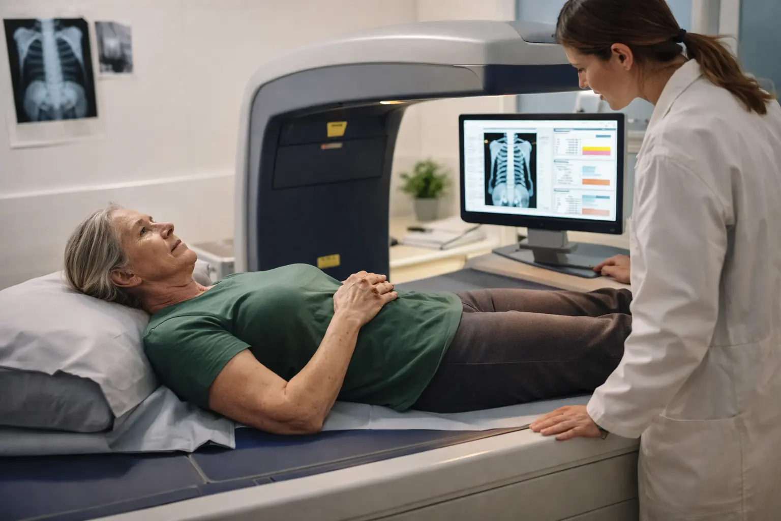
You’re worried about your bones breaking from a simple fall, and nobody’s really explained what’s actually happening inside your skeleton, so let’s cut through the confusion and get you the bone density scan dxa explained in plain English.
Understanding bone density scan dxa
A DXA scan, short for Dual-Energy X-ray Absorptiometry, is a painless and non-invasive test that measures how much mineral is packed into your bones. Think of it like taking a snapshot of your skeletal strength. This scan has become the gold standard for detecting osteoporosis and assessing your personal fracture risk. Imagine you’re 68 years old and you’ve noticed your posture changing slightly, or perhaps a family member experienced a hip fracture from a minor stumble. That’s when your doctor might recommend a DXA scan. The test compares your bone density measurements to established reference standards based on healthy young adults, helping determine whether your bones are in the normal range, showing signs of bone loss (osteopenia), or have progressed to osteoporosis. For seniors, this information becomes crucial because it allows for early intervention before a serious fracture occurs.
The science behind dxa technology
DXA technology operates on a fascinating principle involving two different X-ray beams at distinct energy levels passing through your bones. As these beams travel through your skeletal tissue, the bone absorbs varying amounts of radiation depending on its mineral density. A bone that is denser and stronger will absorb more X-rays, while bones with lower mineral content allow more radiation to pass through. The scanner detects this difference and uses sophisticated software to calculate your bone mineral density, typically measured in grams per square centimeter. Picture yourself lying still on a padded table for about 10 to 30 minutes while the scanning arm moves slowly across your hip, spine, and sometimes your forearm. The entire process is quiet and requires no injections or special preparation. The radiation exposure is minimal, roughly equivalent to the natural background radiation you receive in a single day from the environment. This makes DXA remarkably safe for repeated monitoring over time.
Interpreting the results of a dxa scan
Your DXA results arrive as a T-score and Z-score, which might sound technical but are actually straightforward once explained. The T-score compares your bone density to that of a healthy 30-year-old adult, while the Z-score compares you to people your own age and gender. A T-score of minus 1.0 or higher indicates normal bone density. Scores between minus 1.0 and minus 2.5 suggest osteopenia, meaning your bones are weaker than normal but not yet in the osteoporosis range. A T-score below minus 2.5 indicates osteoporosis. Consider a real scenario: Margaret, a 72-year-old woman, received her DXA results showing a T-score of minus 2.8 in her hip and minus 2.2 in her spine. This meant she had osteoporosis in her hip but osteopenia in her spine, requiring different management strategies for each area. Understanding these numbers helps you and your healthcare provider make informed decisions about treatment and lifestyle modifications.
- Schedule a consultation with your healthcare provider to review your specific T-scores and Z-scores in context of your personal health history and risk factors.
- If your results show low bone density or osteoporosis, discuss treatment options ranging from dietary changes and exercise programs to medications like bisphosphonates or hormone-related therapies.
- Establish a follow-up schedule with your healthcare professional, typically involving repeat DXA scans every 1 to 2 years to monitor whether your bone density is stable, improving, or declining over time.
🔬 Science-backed benefits in 2 minuteschoose where to begin:
Key benefits of dxa scans for seniors
For seniors, DXA scans serve as an early warning system for bone health problems before they become serious. A fracture from osteoporosis can fundamentally change a senior’s life, leading to loss of independence, chronic pain, and reduced quality of life. Hip fractures, in particular, often require surgery and extended rehabilitation, sometimes resulting in permanent mobility loss. By detecting bone density loss early through DXA screening, you gain the opportunity to intervene with targeted treatments and lifestyle modifications before a fracture occurs. Consider the case of Robert, a 75-year-old man who had a DXA scan done routinely and discovered moderate osteoporosis. With this knowledge, he began a structured exercise program, increased his calcium and vitamin D intake, and started medication. Two years later, his follow-up scan showed stabilized bone density, and he remained fracture-free and active. This preventive approach exemplifies how DXA scans empower seniors to take control of their skeletal health and maintain their independence and active lifestyle.
Factors affecting dxa scan results
Your DXA results don’t exist in a vacuum; numerous biological and lifestyle factors influence the measurements your scan produces. Age naturally affects bone density, as bone loss accelerates after age 50, particularly in women following menopause due to declining estrogen levels. Gender matters significantly, with women generally having lower peak bone mass than men and experiencing faster bone loss. Certain medical conditions like rheumatoid arthritis, kidney disease, and thyroid disorders can impact bone density. Medications such as corticosteroids used for conditions like asthma or lupus can weaken bones over time. Your body weight plays a role too, as heavier individuals typically have higher bone density due to increased mechanical stress on bones. Nutritional status, including calcium and vitamin D levels, directly influences bone strength. Lifestyle factors like smoking, excessive alcohol consumption, and sedentary behavior all contribute to lower bone density. When you meet with your healthcare provider before your DXA scan, providing accurate information about your medical history, medications, and lifestyle helps ensure the most reliable interpretation of your results.
Future outlook in bone density assessment
The field of bone density assessment continues to evolve with remarkable technological advances. Researchers are developing enhanced DXA software that can provide more detailed analysis of bone microarchitecture and strength beyond simple mineral density measurements. Artificial intelligence and machine learning algorithms are being integrated into DXA systems to improve fracture risk prediction with greater accuracy. Some facilities are exploring advanced imaging techniques like high-resolution peripheral quantitative CT scanning, which can visualize bone structure in extraordinary detail. Digital health platforms are emerging that allow seniors to track their bone health over time, receive personalized recommendations, and communicate results directly with their healthcare providers. These innovations aim to make bone density assessment more precise, accessible, and actionable. The integration of genetic testing with imaging results may soon allow for truly personalized bone health management strategies. As these technologies mature and become more widely available, seniors will benefit from increasingly sophisticated tools to understand and manage their skeletal health throughout their aging years.
DXA scans offer valuable insights into bone health by providing accurate measurements of bone density. Understanding your DXA results can guide effective interventions and preventive measures to maintain skeletal health as you age.
Are DXA scans safe for seniors?
Yes, DXA scans are considered safe for seniors as they involve minimal radiation exposure, equivalent to a day of natural background radiation.
How often should seniors undergo DXA scans?
The frequency of DXA scans for seniors depends on individual risk factors and bone health status, with recommendations varying from every 1 to 2 years.
Others also read:
Disclaimer: This article is for informational purposes only and is not a substitute for professional medical advice. Always consult a healthcare professional for personal guidance.
This article has been prepared and reviewed by the GlobalHealthBeacon editorial team and is based on current medical research and published scientific literature available in 2026. It provides structured, evidence-based information to support informed health decisions.
