You’re worried about your bones breaking from a simple fall, and nobody’s giving you straight answers about what’s actually happening inside your skeleton – but a bone density scan dxa explained could finally show you exactly where you stand.
What is a DXA bone density scan?
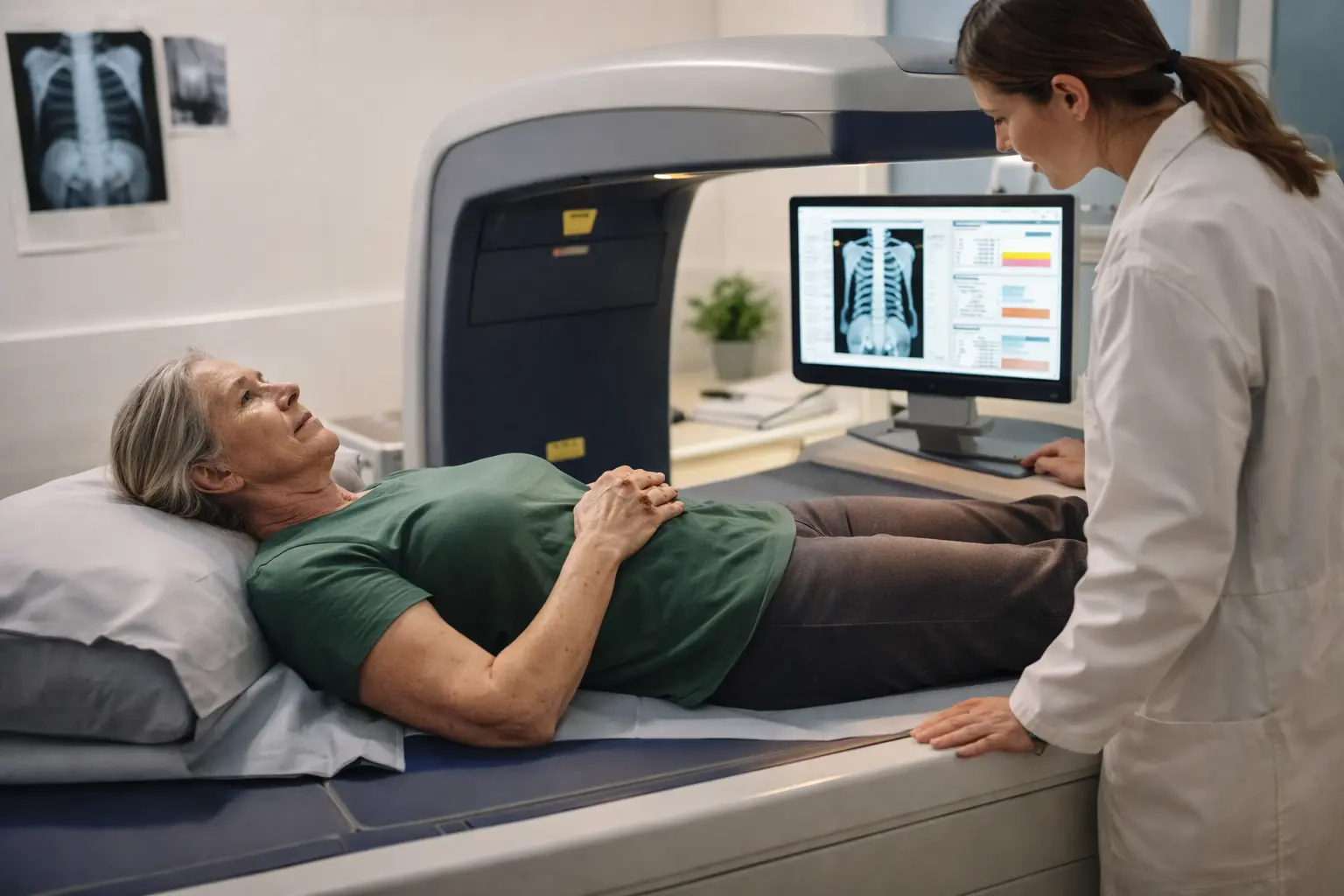
A DXA scan, short for dual-energy X-ray absorptiometry, is a painless and non-invasive imaging test that measures bone mineral density (BMD) by using two different energy levels of X-rays to distinguish bone from soft tissue. Think of it as a specialized photograph that reveals the actual density and strength of your bones compared to a reference standard. The test takes about 10 to 30 minutes depending on which bones are being scanned, typically the hip, spine, and forearm. During the scan, you lie flat on a padded table while a scanning arm passes over your body, creating detailed images without any discomfort or injection. Unlike a regular X-ray that shows bone structure, a DXA scan quantifies exactly how much mineral content your bones contain, which directly correlates to fracture risk and overall skeletal health.
Why is DXA testing important for seniors?
As we age, our bones naturally lose density through a process called bone resorption, where the body breaks down bone faster than it rebuilds it, making them more susceptible to fractures from minor falls or even simple movements. For seniors, a fracture can be life-altering: a hip fracture often requires surgery and months of recovery, potentially leading to permanent mobility loss or loss of independence. DXA scans are crucial in identifying osteoporosis early, sometimes years before a fracture occurs, allowing time for intervention through lifestyle changes or medications. Consider a 68-year-old woman who feels fine but discovers through a DXA scan that her bone density is significantly below normal; this early detection gives her the opportunity to start treatment and prevent a devastating fracture that could have occurred within the next few years. Regular monitoring through DXA scans also tracks whether treatments are working effectively, ensuring your bone health strategy is actually protecting you.
How to prepare for a DXA scan
Preparing for a DXA scan is straightforward, but following these steps ensures your results are accurate and the process goes smoothly. Wear comfortable, loose-fitting clothing without metal zippers, buttons, or snaps because metal interferes with the X-ray imaging and may require you to change into a hospital gown, wasting time. Remove all jewelry, including watches, rings, and necklaces, and let your technician know if you have any metal implants like joint replacements or pacemakers. Inform your doctor about any recent imaging tests involving barium or contrast material, as these can linger in your system and create false readings on your bone density results. Avoid taking calcium supplements for at least 24 hours before your scan because concentrated calcium in your digestive system can artificially inflate your bone density measurements, making your bones appear denser than they actually are. If you take medications, continue them as normal unless your doctor specifically advises otherwise. Arrive a few minutes early to complete paperwork, and plan to be there for about 30 to 45 minutes total.
- Wear comfortable clothing without metal accessories.
- Inform your doctor of any recent scans with barium or contrast material.
- Avoid calcium supplements 24 hours before the scan.
🔬 Science-backed benefits in 2 minuteschoose where to begin:
Understanding your DXA results
After your DXA scan, your doctor will provide you with a T-score, which compares your bone density to that of a healthy young adult at peak bone mass, essentially showing how many standard deviations your bones are above or below this reference point. The T-score scale is straightforward: a T-score of -1 or above is considered normal bone density, meaning your fracture risk is low and no treatment is typically needed. A T-score between -1 and -2.5 indicates low bone mass, sometimes called osteopenia, which means your bones are weaker than normal but not yet at the osteoporosis threshold, though you should discuss prevention strategies with your doctor. A T-score of -2.5 or lower signifies osteoporosis, indicating significantly weakened bones with high fracture risk, requiring medical attention and likely treatment. Your results also include a 10-year fracture probability, which estimates your actual risk of breaking a hip, spine, or other major bone in the next decade. For example, a 70-year-old woman with a T-score of -3.0 might have a 20 percent chance of a hip fracture in the next 10 years, which would warrant serious consideration of treatment options.
Benefits and risks of DXA testing for seniors
The primary benefit of DXA testing is early detection of bone loss before it becomes severe enough to cause fractures, giving you years to intervene through lifestyle modifications, dietary changes, or medications that can slow or even reverse bone loss. DXA scans also help identify which seniors are at highest risk, allowing doctors to prioritize treatment for those who need it most and avoid unnecessary medication for those with minimal risk. The test provides objective data rather than guesswork, so you and your doctor can make informed decisions based on actual bone density measurements rather than assumptions. The radiation exposure from a DXA scan is minimal, roughly equivalent to the radiation you receive during a short airplane flight or a few hours in the sun, making it extremely safe even with repeated scans over many years. There are virtually no contraindications or side effects from the procedure itself. The main limitation is that DXA measures bone density but not bone quality or architecture, so two people with identical T-scores might have different fracture risks based on other factors like balance, muscle strength, or previous falls.
Interpreting DXA results with your doctor
Your DXA results are just the beginning of a conversation with your healthcare provider about your bone health strategy and future fracture prevention. Bring your results to your appointment and discuss not just the numbers but what they mean for your specific situation, considering your age, gender, medical history, medications, and lifestyle factors. Your doctor will explain whether your results warrant treatment, and if so, what options are available, ranging from lifestyle changes like weight-bearing exercise and adequate calcium and vitamin D intake to prescription medications like bisphosphonates that slow bone loss. If you have osteoporosis, your doctor might recommend fall prevention strategies such as home safety assessments, vision and hearing checks, and balance training, since preventing falls is just as important as strengthening bones. Discuss how often you should have repeat DXA scans, typically every one to two years if you have osteoporosis or are on treatment, to monitor whether your bone density is stabilizing or improving. Ask about any medications you’re currently taking that might affect bone health, and whether your diet provides adequate calcium and vitamin D, or if supplements are needed.
DXA scans are essential for seniors to assess bone health and osteoporosis risk with scientific accuracy and minimal radiation exposure. Understanding your T-score results and working closely with your healthcare provider to develop a personalized bone health plan can help maintain bone density, reduce fracture risk, and preserve your independence and quality of life as you age.
Is a DXA scan safe for seniors?
Yes, DXA scans are safe and painless, exposing you to minimal radiation levels comparable to a short flight or a sunny day. The procedure has no side effects, no needles, and no contrast dye, making it one of the safest diagnostic tests available for seniors.
How often should seniors have a DXA scan?
Seniors with normal bone density may need a DXA scan every 2 years, while those with osteoporosis or low bone mass may require more frequent monitoring, typically annually or every 18 months, as advised by their healthcare provider based on their individual risk factors and treatment response.
Others also read:
Disclaimer: This article is for informational purposes only and is not a substitute for professional medical advice. Always consult a healthcare professional for personal guidance.
This article has been prepared and reviewed by the GlobalHealthBeacon editorial team and is based on current medical research and published scientific literature available in 2026. It provides structured, evidence-based information to support informed health decisions.
