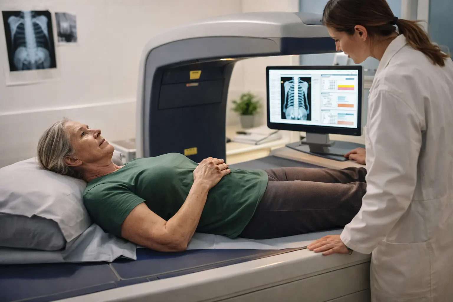
You’re worried your bones are getting weaker, you’ve heard about osteoporosis, and now your doctor mentions a bone density scan DXA explained – but nobody tells you what actually happens or what those confusing numbers mean, so let’s cut through the confusion right now.
What is a bone density scan DXA?
A Bone Density Scan DXA, also known as Dual-energy X-ray absorptiometry, is a non-invasive test that measures bone mineral density with precision. Think of it like a detailed report card for your skeletal system. The scan uses low-dose X-rays to evaluate how dense your bones are, particularly in high-risk areas like your hip and spine where fractures tend to happen. Imagine your bones as a structure: when you’re younger, they’re tightly packed and strong. Over time, especially after age 50, bones naturally lose density and become more porous, like a sponge losing its firmness. This test catches that process early. It’s commonly used to diagnose osteoporosis, a condition where bones become brittle and fracture easily, and to assess your personal fracture risk. The procedure takes less than 15 minutes and exposes you to less radiation than a cross-country flight. For seniors, this scan is often the first real look at whether bone loss is happening and how serious it might be.
- Helps in identifying osteoporosis and assessing fracture risk
- Uses low-dose X-rays to measure bone mineral density
- Commonly focuses on hip and spine regions
Preparing for a bone density scan DXA
Getting ready for your DXA scan is straightforward, but a few details matter. Start by telling your healthcare provider about any medical conditions you have, especially thyroid problems, kidney disease, or anything that affects calcium absorption. List all medications and supplements you’re taking, as some can influence bone density readings. Wear loose, comfortable clothing without metal zippers, buttons, or snaps because metal interferes with the X-ray images. Leave jewelry, watches, and belts at home. About 24 hours before your appointment, skip calcium supplements and calcium-fortified foods if possible, as they can create a false reading. You don’t need to fast or change your normal routine otherwise. If you’ve had a recent CT scan or nuclear medicine test with contrast dye, mention this to your technician because it can affect results. Arrive 10 minutes early to complete paperwork. Think of this preparation like tuning an instrument before a performance: small adjustments ensure accurate results.
📘 Fix your day in under 2 minuteschoose where to begin:
What to expect during the procedure
Walk into the scanning room and you’ll see a table that looks similar to an X-ray bed, with an arm suspended above it. The technician will position you on your back and ask you to lie very still. You might feel a bit vulnerable or anxious, which is completely normal. The scanning arm slowly passes over your body, emitting low-dose X-rays while a detector below the table captures the images. You won’t feel anything during this process, hear much noise, or experience any discomfort. The whole scan typically takes 10 to 15 minutes. The technician might ask you to hold your breath briefly during certain scans to reduce movement blur. You can ask questions anytime. Most facilities have a monitor where you can watch the process, which helps some people feel less anxious. Once finished, you’re done. No recovery time needed. You can return to your normal activities immediately, eat normally, and resume any medications. Results are usually available within a few days.
Understanding the results
Your DXA results arrive as a report with two key numbers: the T-score and the Z-score. The T-score compares your bone density to that of a healthy 30-year-old of your same gender. A T-score of -1.0 or higher is considered normal. Between -1.0 and -2.5 means you have low bone mass, often called osteopenia. Below -2.5 indicates osteoporosis. The Z-score compares you to people your own age and gender, which gives context about whether your bone loss is typical for your age or more severe. For example, a 75-year-old woman might have a T-score of -2.8, indicating osteoporosis, but a Z-score of -0.5, meaning her bone density is actually better than average for her age group. Your healthcare provider will review both scores and discuss what they mean for your fracture risk. They’ll consider your overall health, fall risk, family history, and lifestyle. This conversation is where the real guidance happens. Don’t hesitate to ask your doctor to explain anything that feels unclear.
Benefits of a bone density scan DXA
Early detection of bone loss is genuinely life-changing. When osteoporosis is caught before a fracture happens, you have time to act. You can start strength training, adjust your diet, take medications if needed, and make home modifications to prevent falls. A hip fracture in a senior often leads to surgery, months of recovery, loss of independence, and sometimes permanent disability. A spine fracture can cause chronic pain and loss of height. The DXA scan gives you a window to prevent these outcomes. Beyond prevention, the scan provides a baseline measurement. If you’re already on osteoporosis treatment, repeat scans every two years show whether your medication is working. This feedback loop is powerful. You’re not guessing whether your efforts matter; you have data. The scan also identifies which bones need the most attention. If your hip is strong but your spine is weak, your doctor can tailor recommendations. For seniors, this test is a vital tool in maintaining independence, reducing fracture risk, and staying active in the years ahead.
A Bone Density Scan DXA, or Dual-energy X-ray absorptiometry, is a non-invasive test that measures bone mineral density, helping to diagnose osteoporosis and assess fracture risk. Preparing for the scan, understanding the procedure, interpreting the results, and realizing the benefits are crucial steps in taking control of your bone health.
How often should seniors get a bone density scan DXA?
It is recommended that women aged 65 and older and men aged 70 and older undergo a bone density scan DXA. Your healthcare provider may suggest earlier testing based on your risk factors.
Are there any risks associated with a bone density scan DXA?
A Bone Density Scan DXA is a safe procedure with minimal radiation exposure. The benefits of early detection of osteoporosis and fracture risk outweigh the risks associated with the scan.
Others also read:
Disclaimer: This article is for informational purposes only and is not a substitute for professional medical advice. Always consult a healthcare professional for personal guidance.
This guide has been prepared and reviewed by the GlobalHealthBeacon editorial team and reflects current medical research as of 2026. It provides structured, evidence-based information to support informed health decisions.
